ENGR2019AGUEROADAME49081 ENGR
Engineering Capstone Project - Computer Vision
Type: Undergraduate
Author(s):
Melina Aguero Adame
Engineering
Susana Murillo
Engineering
Advisor(s):
Stephen Weis
Engineering
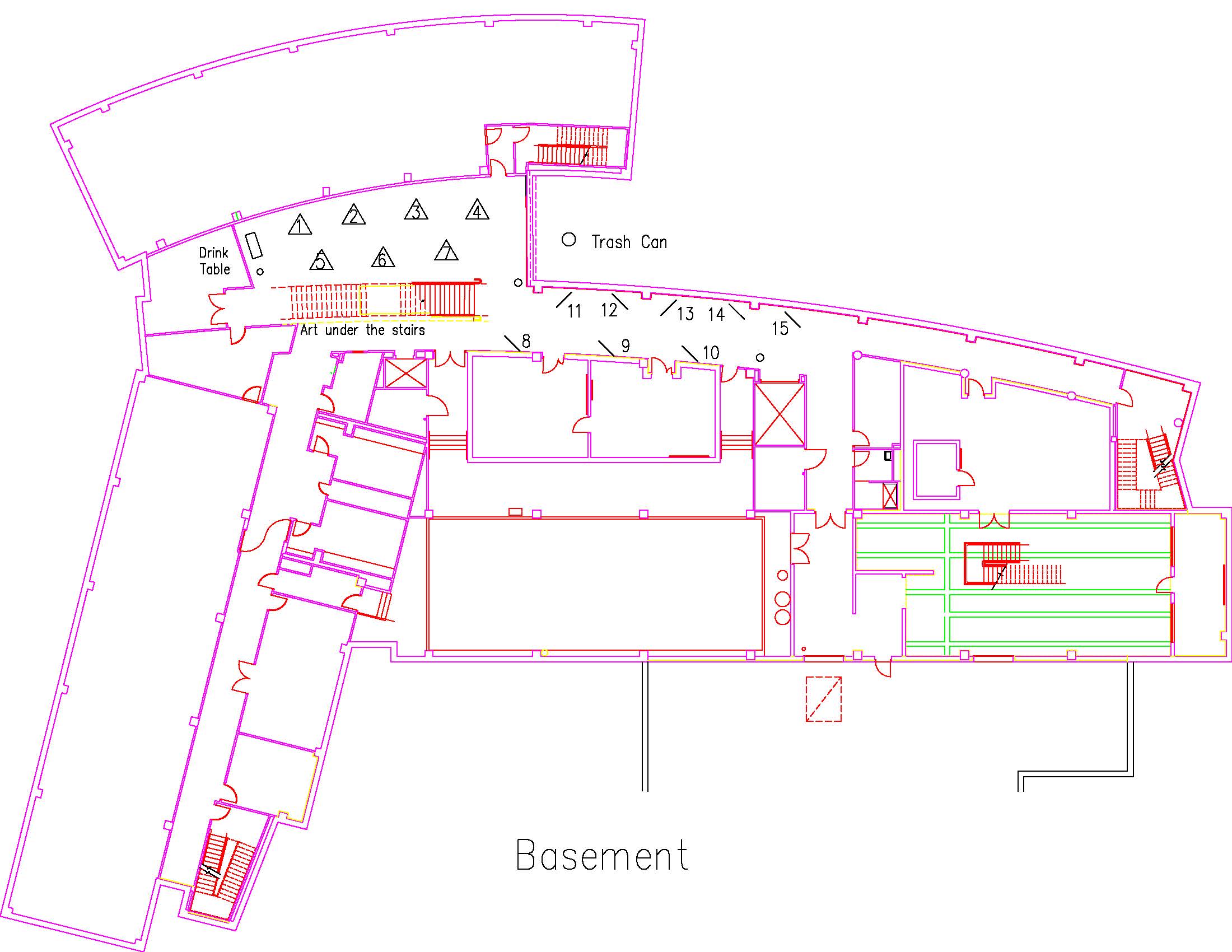
Location: Session: 1; Basement; Table Number: 6
View PresentationAs part of one of the engineering capstone projects, a calibration testing system was improved with the aid of computer vision. Computer vision was integrated into this project as a solution to a rotating pedestal calibration test that was previously performed by the naked eye. The main goal of this system was to detect and track a red 635 nm wavelength laser spot with offsets as small as 0.025 inches on a 10 x 10 inch grid accurately and precisely. Designing this system involved three major criteria: camera selection, data processing hardware, and algorithm performance.
The first criteria studied in the design process was the camera. The system required a camera that was compact in size, covered the entirety of the grid at less than 11 inches, and captured high quality images. Furthermore, two main data processing hardwares were explored: Raspberry Pi and a standard test laptop. The processing hardware criteria considered were speed, portability, and maintenance. Finally, RGB and houghcircles were the two algorithms used to detect the red laser dot. Testing was conducted to compare the algorithms based on their ability to detect the laser spot, precision in tracking, and repeatability. These design considerations guided the down selects for the final components used in this system.
ENGR2019BIESEMEIER44452 ENGR
Design and Development of an Actuation and Extraction Force Tester: Programming
Type: Undergraduate
Author(s):
Thomas Biesemeier
Engineering
Zach Hollis
Engineering
Ben Krause
Engineering
Talha Mushtaq
Engineering
Advisor(s):
Robert Bittle
Engineering
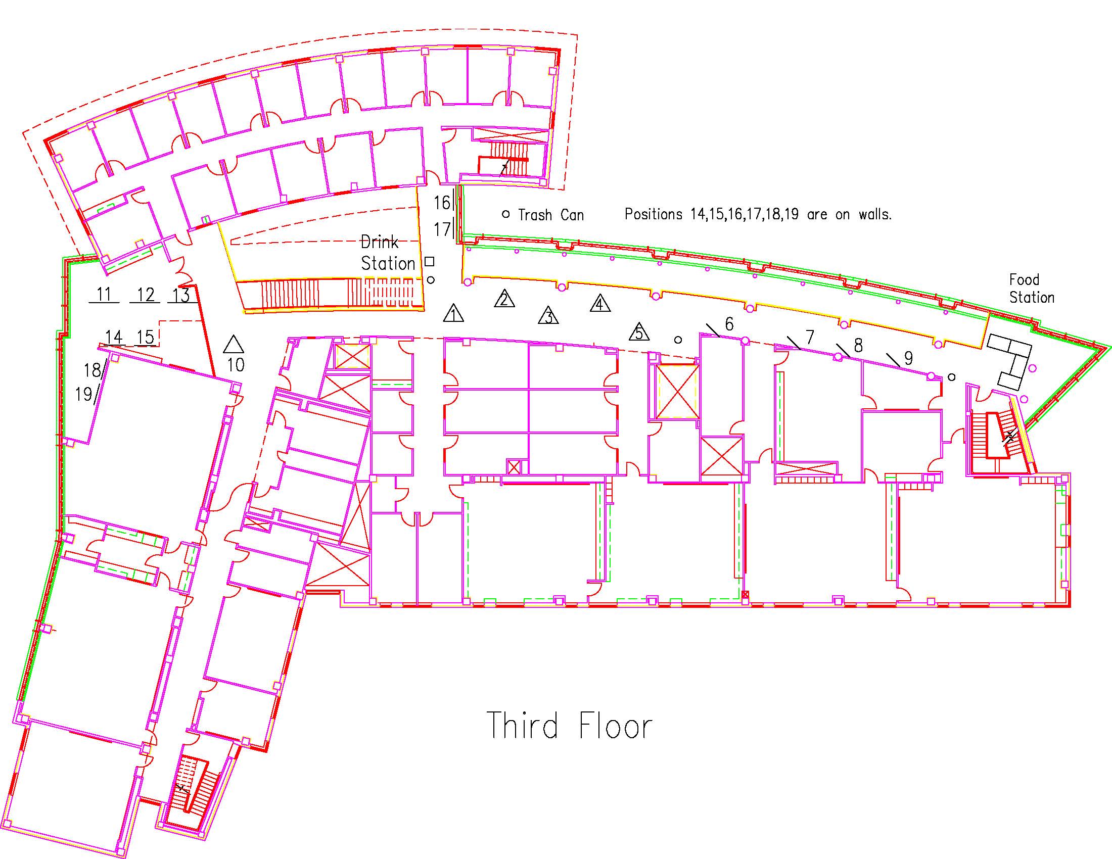
Location: Session: 2; 3rd Floor; Table Number: 3
View PresentationThe LabVIEW team for the Applied Avionics Inc. project focuses on fully integrating the programming of all electrical components with LabVIEW. The major requirements for this project include utilizing LabVIEW to display and capture data feedback, completely automate the testing process, and to read and send data directly to AAI’s database. By creating an actuation and extraction feedback machine that is fully LabVIEW controlled, a variety of switch body types were able to be accommodated and tested. The machine has been shown to decrease variability of results and improve the efficiency of AAI’s current process in all aspects required.
ENGR2019CLARKE58037 COSC
AWS for HealthCare
Type: Undergraduate
Author(s):
Kenzie Clarke
Computer Science
Kien Nguyen
Computer Science
Advisor(s):
Cuiling Gong
Engineering
Liran Ma
Computer Science
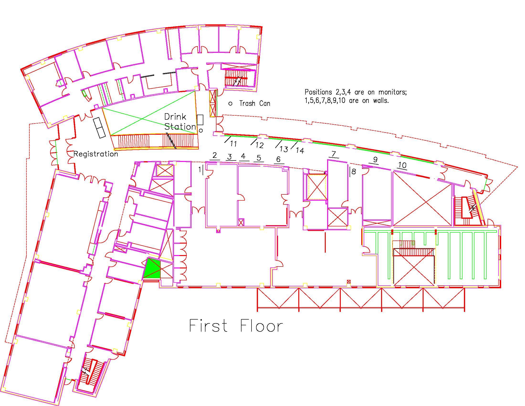
Location: Session: 1; 1st Floor; Table Number: 4
View PresentationCloud based services such as IBM Cloud and Amazon Web Services provides a new platform for data collection, storage and processing through the internet that enables environment monitoring via wireless sensor networks. In this project, we would like to develop a cloud-based low power monitoring and notification platform using AWS. Most existing notification platforms are provided as an expensive, closed system that do not allow flexibility and is often difficult to troubleshoot. These systems require special hardware (such as unique walkie-talkies) and upgrades are pushed back due to costs.
Our system will utilize AWS Lambda functions, a cloud database, and IOT buttons so that medical staff can receive and store real time patient vitals and notifications with a data forwarding device such as a smart phone, tablet, or computer. AWS solutions are low-cost and flexible, allowing the care centers to customize the functionality to their specific needs. These buttons do not require wired power supply and have a long-lasting battery.
ENGR2019DECK64177 ENGR
HyPIR Electrolysis for Potassium Hydroxide Solutions at Different Laser Specifications
Type: Undergraduate
Author(s):
Trystan Deck
Engineering
Aliesha Rau
Engineering
Advisor(s):
John Fanchi
Engineering
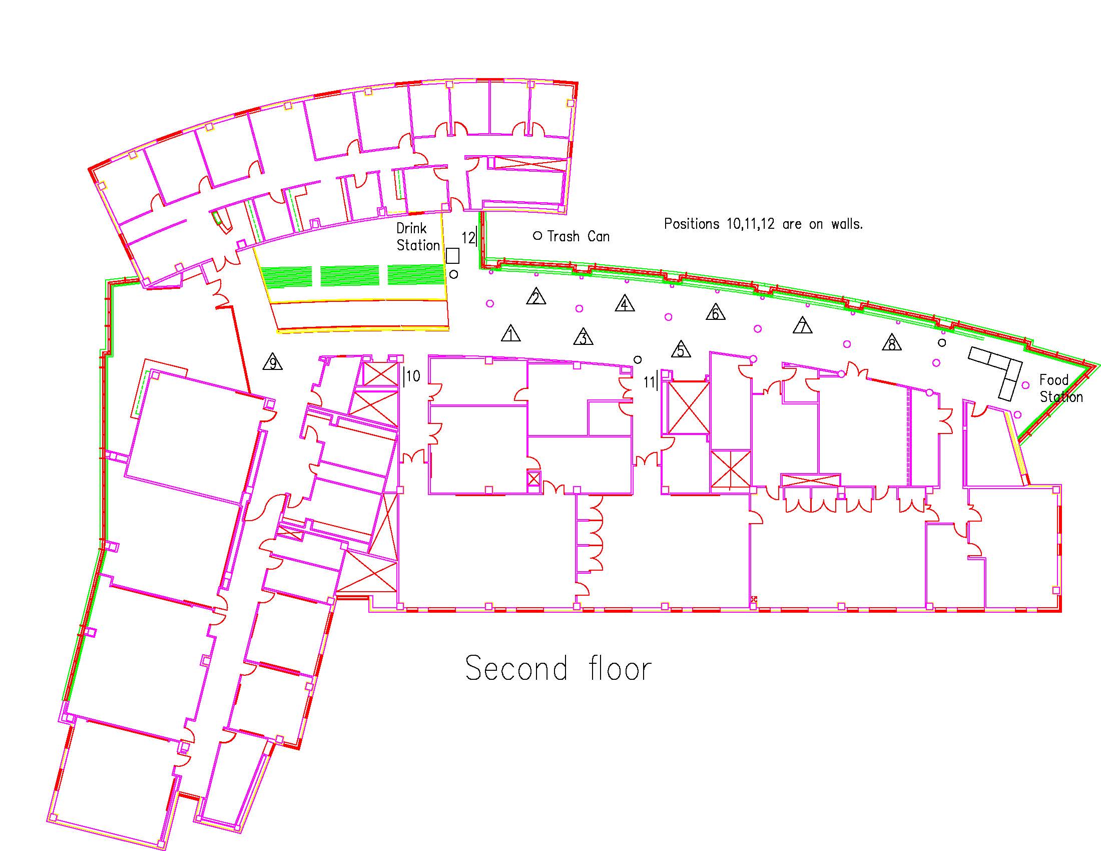
Location: Session: 2; 2nd Floor; Table Number: 9
View PresentationWe are presenting a method referred to as Hydrogen Production by HyPIR Electrolysis. The method increases the rate of hydrogen production from a 1 molar potassium hydroxide and water solution under 6 volts when an infrared laser is irradiated with an optimum wavelength of light through a cell and concentrated on exposed copper electrodes. The irradiating light facilitates the dissociation of water by stretching the hydrogen oxygen bonds and increasing the rate of hydrogen production. Production of hydrogen due to the class 4 laser is altered by the specifications of laser energy, pulses per second, and spot size.
ENGR2019DEVOOGHT49649 ENGR
FDM 3D Printing Mechanical Property Testing
Type: Undergraduate
Author(s):
Luke Devooght
Engineering
Melina Aguero
Engineering
Advisor(s):
Becky Bittle
Engineering
Location: Session: 2; Basement; Table Number: 6
View PresentationIn this experiment, the mechanical properties of 3D printed specimens of different printing parameters were tested under tension. The printing parameters of these specimens were: surface resolution, infill density, and print orientation. Parts were printed in Onyx nylon with a Fused Deposition Modeling (FDM) printer called the Markforged Onyx Pro. Factorial sets of specimens using all various parameters are printed and tested to create a reference table for future engineering projects. Specimens are then printed as composite variations with continuous fibers in order to understand the benefits a composite may have.